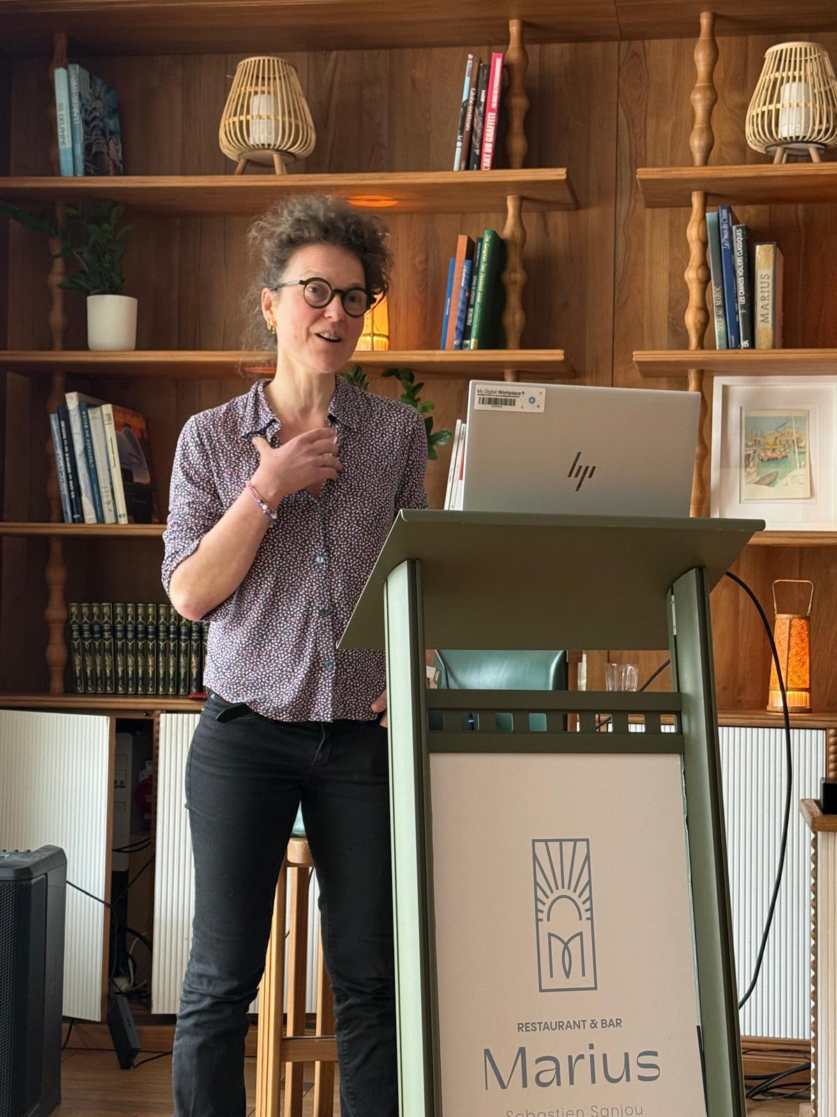
With Dr. Sarah Watson from Institut Curie, Société Générale opens a decisive debate on the future of care
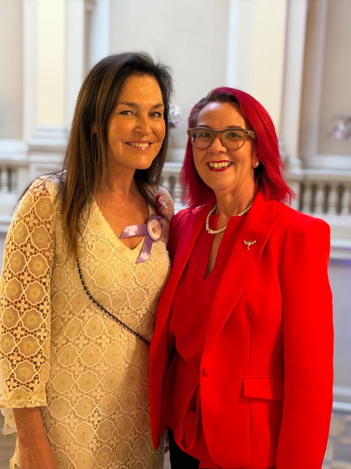
In Monaco, before Société Générale’s women’s club, Dr. Sarah Watson, medical oncologist and researcher at Institut Curie, offered a concrete perspective on artificial intelligence in healthcare. Far from vague promises, she demonstrated how these tools are already transforming diagnosis, guiding treatments, and improving, in certain cases, the survival of patients with particularly complex cancers. A dense, lucid presentation that repositions AI not on the side of technological fantasy, but on the side of clinical decision-making.
In debates about artificial intelligence, superlatives often move faster than evidence. Revolutions are promised, fears are stirred, radical disruptions are announced. But when a physician-researcher speaks from the frontlines of cancer care, the perspective changes. Abstraction gives way to experience. Technology becomes a tool once again. And the central question returns to this: at what point does innovation actually improve patient care?
This is precisely the shift that Dr. Sarah Watson achieved during a conference organized in Monaco by Société Générale for its women’s club. The conference was part of both a relational and societal dynamic. The organizers emphasized their commitment to fostering a community of exchange and reflection. They also highlighted the bank’s support for Institut Curie, particularly through campaigns linked to Breast Cancer Awareness Month. But very quickly, the essential emerged: talking about cancer, talking about research, talking about what AI is already changing in medicine.
A credible voice, rooted in care
Dr. Sarah Watson does not speak from a distant observatory of innovation. At Institut Curie, she divides her time between clinical practice and research. As a medical oncologist, she treats patients with cancers, particularly rare and aggressive forms. As a researcher, she leads a team working on developing artificial intelligence tools applied to oncology.
This dual position gives her discourse particular strength. She knows both the promises of algorithms and the constraints of hospital reality. She knows what a model can provide. She also knows what it doesn’t see, what it doesn’t understand, what still needs interpretation. This is undoubtedly what gave her conference its accuracy. It was neither about selling AI nor fearing it on principle, but evaluating it against what it already enables in the patient journey.
The introduction devoted to AI’s history and its major definitions was briefly mentioned. Yes, artificial intelligence doesn’t date from 2022. Yes, machine learning and deep learning correspond to distinct levels of algorithmic sophistication. Yes, everything rests on two fundamental levers: data and computing power. But the interest of the presentation lay elsewhere. It resided in this infinitely more concrete question: what can AI do when a physician must diagnose cancer, understand its origin, or choose a treatment?
Cancer, a disease that produces data at every stage
One of the most enlightening contributions of the conference was showing that oncology has become a privileged terrain for AI because it generates a considerable mass of data. Even before diagnosis, there is prevention and screening data: mammograms, imaging examinations, medical history. At the moment of diagnosis, radiological images, pathology slides, medical reports, and molecular tumor profiles are added. During treatment, therapeutic responses, side effects, and evolutionary trajectories are further layered. Then, in follow-up, new data appears.
In other words, cancer leaves measurable traces everywhere. And this is precisely where artificial intelligence becomes interesting. It excels when it comes to identifying patterns, detecting correlations, classifying forms, and predicting evolutions from large volumes of information. This is not intelligence in the human sense of the term. It is a statistical and computational capacity to find, in the noise of data, signals that the human eye or mind do not always perceive.
Seeing what the physician doesn’t always see
The first family of applications presented concerns image analysis. In oncology, this dimension is central. A tumor is read in a mammogram, in a CT scan, in a PET scan, in a histological image. However, these images can now be analyzed by models trained on thousands, sometimes hundreds of thousands of cases.
Dr. Watson recalled that a first turning point had been reached in dermatology. Tools have learned to distinguish benign lesions from cancerous ones from skin images. In certain studies, these systems have surpassed the performance of experienced dermatologists. The issue is not to disqualify the specialist. It is to understand that a machine, when well trained, can become a powerful aid in detecting early signals, standardizing interpretations, and reducing certain errors.
Mammography is another major example. Here, AI’s value is twofold. It can improve the detection of already present cancer, but also help estimate the risk that cancer will appear in the following years from images deemed normal. The prospect is considerable. It opens the way to less uniform, therefore more personalized screening. Instead of applying the same surveillance rhythm to all women, one could imagine pathways modulated according to actual risk. Some would be reassured and monitored more lightly. Others, identified as more exposed, would benefit from closer monitoring.
This shift is not trivial. It touches on a sensitive question: how to move away from mass medicine without falling into opaque medicine? Because personalizing also means explaining. And this is where one of the major tensions of medical AI appears: a tool can be effective without being immediately intelligible to those who use it.
Anatomical pathology enters a new era
One of the most interesting passages of the conference concerned anatomical pathology, the field where tissues taken from patients are observed under a microscope to confirm and characterize cancer. What Dr. Watson emphasized is essential: a histological image is not just a photograph. It is a concentration of biological information. In the organization of a tumor, in cellular architecture and in certain textures invisible to classic human analysis, the machine can detect relevant signals.
They provide information about the disease’s aggressiveness, the risk of recurrence, sensitivity to treatments, or certain genetic characteristics.
We are entering here into medicine augmented by inference. From a simple image, the algorithm does not only identify what is there. It suggests what could happen. It no longer just reads the tumor’s present. It begins to project its probable future. For medical teams, this potentially means earlier decisions, better calibrated therapeutic choices, and enhanced capacity to stratify risks.
The most striking case: cancers of unknown primary
But it was in addressing cancers of unknown primary that the presentation took on its full significance. These cancers count among the most feared situations in oncology. The patient arrives with metastases. The cancer has therefore already spread. Yet, despite biopsies, imaging, and complementary examinations, physicians cannot identify the organ of origin. We know the disease is there. We see its consequences. But we ignore its starting point.
This situation does not merely constitute intellectual frustration. It entails major therapeutic consequences. In oncology, treatments remain largely determined by the initial tumor type. Each cancer responds to specific logics. When the origin remains unknown, care becomes empirical. Broad-spectrum treatments are administered, with often limited effectiveness and an unfavorable prognosis.
Dr. Watson recounted the case of a 32-year-old patient referred to Institut Curie with numerous metastases visible on PET scan. The first clinical hypothesis suggested sarcoma. The biopsy ultimately ruled out this lead, without providing a clear answer. The pathology report suggested several possible origins without allowing a determination. For the oncologist, this is a dead end. The disease is progressing. Time matters. But the tumor’s identity remains elusive.
When molecular signature becomes a compass
This is where Dr. Watson’s team formulated a decisive hypothesis. Even when cancer becomes metastatic and seems to have lost its initial appearance, it perhaps retains a memory of its organ of origin. Not in its visible form, but in its molecular signature. From there, the reasoning became experimental: train an AI on thousands of genetic profiles of known cancers, then submit tumors of unknown origin to see if it can recognize their signature.
The project mobilized more than 20,000 molecular samples from European and American databases. Initially, the team let the machine work in unsupervised mode. No labels were provided. It had to group profiles according to their similarities on its own. The result was striking: samples spontaneously organized themselves into tumor families. This meant that the molecular signatures of major cancer types were sufficiently distinct to emerge on their own in the algorithmic space.
In a second phase, the tool was applied to cancers of unknown primary. In approximately 80% of cases, it was able to propose a probable origin. For the patient presented during the conference, the machine concluded, with a high score, that it was kidney cancer. This prediction immediately changed the therapeutic strategy. Metastatic kidney cancers respond poorly to certain classic chemotherapies. Following the standard schema for unknown primaries would therefore likely have led to failure. The team chose to embrace the AI’s prediction and treat it as kidney cancer. The patient achieved a prolonged complete response, then survival of several years, where life expectancy had been a few months.
What matters most is not the spectacular effect of the individual case. What matters most is the logic it reveals. AI does not replace medical reasoning. It reorients it. It opens a hypothesis that clinical examination alone had not allowed to stabilize. It makes a more specific decision possible. It transforms uncertainty into a course of action.
From laboratory to multidisciplinary consultation meeting
The second merit of this work is its transition from laboratory to care. Too many AI projects stop at the scientific publication stage. This one has been integrated into a national multidisciplinary consultation meeting dedicated to cancers of unknown primary. In other words, the algorithm did not remain a prototype. It became a decision support tool in a real clinical organization.
The results presented are significant. In more than 70% of cases, using this approach modifies the retained diagnosis. And when treatment is guided based on the AI’s prediction, overall median survival increases noticeably. Dr. Watson cited a shift from 11 to 19 months. These figures should not be over-interpreted. They do not mean that AI cures incurable cancers. But they indicate that it can concretely improve the quality of therapeutic guidance. In oncology, where every month gained has weight, this progression is far from marginal.
The challenges are now political, organizational, and human
This conference was not a naive plea for unbridled automation. Dr. Watson insisted on the limitations and obstacles. The first relates to data. In France and Europe, it exists, but remains poorly harmonized, difficult to share, locked in heterogeneous information systems. The second relates to evaluation. Too many tools emerge without a sufficiently robust framework to compare their performance, verify their validity on independent datasets, and guarantee their actual safety. The third challenge is human: physicians are not yet trained to work with these tools.
The remark is important. The problem is not only technological. It is cultural. Practitioners must learn to read the results produced by these systems, understand their blind spots, discuss their recommendations. Responsibilities must also be clarified. If AI suggests an erroneous strategy, who answers? The practitioner, today, remains legally at the center. This assumes that AI remains a support, not a substitute.
More human medicine thanks to machines?
The conclusion of the presentation, perhaps the strongest, addressed a paradox. Certain studies comparing physicians’ and chatbots’ responses to patient questions show that conversational systems are sometimes judged more empathetic. Should we conclude that the machine is becoming more human than the caregiver? Dr. Watson had the intelligence to temper this observation. A chatbot is never tired. It doesn’t end its day after hours of consultations, files, emergencies, difficult announcements. It always responds with the same apparent availability.
But the real lesson lies elsewhere. If AI takes charge of repetitive tasks, facilitates data synthesis, and accelerates the guidance of complex decisions, it can free up medical time.
This time can be reinvested in what remains essential: attention given to the patient. Time to look, to explain, to accompany, to doubt with the patient, to fully exercise that human discernment that no algorithm replaces.
In Monaco, that day, artificial intelligence was not presented as a myth of power. It appeared for what it is becoming in oncology: a precision instrument in situations where medicine alone sometimes confronts its limits. This does not diminish the physician. It makes their responsibility even more demanding. The future will probably not oppose machine to caregiver. It will rather distinguish two practices: one that will know how to integrate AI with rigor, and one that will remain distant from an already engaged transformation. In medicine, as often, innovation only makes sense when it serves the accuracy of the gesture and the dignity of care.